The Retinacular system of the hand consists of extensor and flexor retinaculum, juncturetendinum, sagittal bands, spiral…
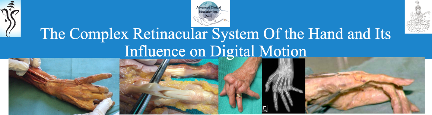
The Retinacular system of the hand consists of extensor and flexor retinaculum, juncture
tendinum, sagittal bands, spiral oblique retinacular ligament, transverse retinacular ligament,
triangular ligaments, and the palmar pulleys. These structures have an intricate relationship with
each other. The importance of these structures on the biomechanics of the digital motion is well
described by various authors including: Tubiana, Bunnell, Brand, and Kapandji. These structures
transmit precise forces during flexion, extension and opposition of the digits, by regulating
excursion of flexors, extensors and the intrinsics for digital function. Extensor or the flexor
tendon injuries, attenuation or adaptive shortening of any of the retinacular structures disrupt the
kinematic chain equilibrium and lead to various hand deformities such as swan neck and
boutonniere/pseudo-boutonniere. Recognition of pathologies, and conducting special clinical
tests directs precise management of the various clinical conditions. Once diagnosed, treatment
may consist of relative motion splinting and standard therapeutic measures to increase joint
motion, tendon excursion, and function.
The presentation includes illustrations, animations clinical and the cadaveric dissection videos to
simplify understanding of the normal and disturbed digital mechanics. The use of the above-
mentioned media is to contribute to the evidence of clinical management.
Course Objectives
Upon completion of the webinar the participants will have: